Articles
Information for Parents Carers of Children Having Investigations in Relation to Unexplained Injuries + Consent form Bruising and Injuries in Babies and Children – Parents Leaflet Multi-Agency Protocol for Injuries to Non-Mobile Children Flowchart for children attending Galloway Community Hospital (GCH) for NAI Follow Up Skeletal Survey Flowchart for children attending DGRI for NAI Follow-Up Skeletal Survey Cognitive Function Conscious Level Kidney Biopsy Complications Parenteral Iron for Non-HD CKD Patients Fracture Management Guidelines (Paediatric) Fracture Management Guidelines (Adult) Management of Hypertension in Acute Stroke Prescribing for CAU Patients Still in ED Hypothermia Deactivation of Implantable Cardioverter Defibrillator Myeloma Croup Care Of Burns In Scotland (COBIS) Paediatric Guidance Management of Epistaxis Sore Throat Differential Diagnosis Dizziness Differential Diagnosis Peritonsillar Abscess/Quinsy Acute Tonsillitis Acute Mastoiditis Otitis Media Otitis Externa Extravasation of IV Amiodarone WoS Paediatric Drooling and Aspiration Guideline Palliative Care – How to Refer Eating Disorders Stroke Care Warfarin Anticoagulation for AF, DVT and PE Molnupiravir MyPsych Foundation Doctors Toolkit Paediatric Febrile Neutropenia Guidance PAEDIATRIC HYPOGLYCAEMIA MANAGEMENT in NON DIABETIC CHILDREN   Paediatric Diabetic Ketoacidosis (DKA) Guideline Child Protection Policies and Procedures (D&G) Management of Acute Behavioural Challenges in Adolescents and Young People presenting to Secondary Care Cancer of Unknown Primary Patients Returning from Interventional Cardiac Procedure Treatment of Malaria Discharging Patients on High Dose Steroids Sotrovimab Paediatric Ketone Correction Guideline Insulin Correction Factor Table (Paediatrics) Management of uncomplicated Henoch-Schonlein Purpura (HSP) in under 16s Management of Hypoglycaemia in Children with Type 1 Diabetes Newly diagnosed diabetic – not in DKA (Walking wounded) Proton Pump Inhibitor Guideline for Neonatal and Paediatrics Stroke – Post Thrombolysis Neonatal Guidelines Gentamicin Prescribing (Paediatrics) Management of Anaphylaxis (Paediatrics) Management of Prolonged Seizures (Convulsive Status Epilepticus) in Children Bronchiolitis Acute Wheeze or Asthma in Paediatrics Conscious Proning Covid-19 Basics Remdesivir Thromboprophylaxis Identifying Patients in the Highest Risk Groups Steroids for Patients with Covid-19 Infection IL-6 Inhibitors – Tocilizumab or Sarilumab Baricitinib Paxlovid (Nirmatrelvir/Ritonavir) Influenza A Inhalers for Adults with Asthma Standard Operating Procedure for AMU Trigger Finger/Thumb Osteoarthritis of the Hand/Thumb Mallet Finger Ganglion Dupuytren’s Contracture De Quervain’s Tenosynovitis Carpal Tunnel Syndrome Prescribing Advice on Admission – Clozapine Prescribing Advice on Admission – Methadone/Buprenorphine Prescribing Advice on Admission – Corticosteroids Prescribing Advice on Admission – Items Not Prescribed by GP Prescribing Advice on Admission – Patients on Chemotherapy Regimes Prescribing Advice on Admission – Medication for Parkinson’s Disease Prescribing Advice on Admission – Insulin Prescribing Advice on Admission Medical Emergencies in Eating Disorders (MEED) Gentamicin & Vancomycin HIV Testing Guidelines Metabolic Syndrome Associated Fatty Liver Disease (MAFLD) Greener Inhaler Prescribing C4 Predischarge Beds Handover Safe and Secure Handling of Medicines Blood Glucose & Steroids IV Fentanyl & Morphine for Acute Pain in Adults Assessment & Management of Acute Pain Hospitalised and Has Coronavirus19 Infection (No suspected Viral Pneumonia Syndrome) Hospitalised Due to Coronavirus19 with Likely Viral Pneumonia Bi-Level NIV S/T Guidelines for CCU Phase Bi-Level NIV S/T Guidelines for ED Phase Adults With Incapacity Premenstrual Syndrome Pelvic USS Boarding Coeliac diagnosis pathway (Adults) Voice clinic Ear Wax Dermatology Squamous Cell Carcinoma (SCC) Malignant Melanoma Basal Cell Carcinoma (BCC) Nipple Discharge Early Cancer Diagnostic Clinic (ECDC) Genetics Referrals Breast Infections Breast Pain Primary Care Prescribing Guidelines Emergency Department Anaesthetics and Chronic Pain Team Respiratory Referrals Chronic Cough Pathway GP Clinical Handbook Test Paediatric Bronchiolitis Early Cancer Diagnosis Clinic (ECDC) Obstetrics & Gynaecology/Medicine Admission Agreement Idiopathic Intrancranial Hypertension Urology Out of Hours Urology Out of Hours Sengstaken/Minnesota Tube for Bleeding Varices Eradication of Helicobacter pylori Transfer from Galloway Community Hospital Repatriation of Patients from Tertiary Hospitals THROMBOPROPHYLAXIS IN PREGNANCY – Appendix 1, Risk factors THROMBOPROPHYLAXIS IN PREGNANCY – Appendix 3, Postnatal assessment & management THROMBOPROPHYLAXIS IN PREGNANCY – Appendix 4 THROMBOPROPHYLAXIS IN PREGNANCY – Appendix 2, Management of women with previous VTE THROMBOPROPHYLAXIS IN PREGNANCY, LABOUR AND THE PUERPERIUM Orthopaedic VTE Risk Assessment Sodium Glucose Transporter 2 Inhibitors (SGLT2i) Cardiology Referrals Vascular Referrals ‘Watershed’ Conditions Myasthenia Gravis Gentamicin in Renal Replacement Therapy Vancomycin in Renal Replacement Therapy REDMAP Poster Realistic Conversations Summary Plan for Deteriorating Health Treatment Escalation Plans Ambulatory Care for Blood and/or Iron Infusion Principles for Light Touch Patients – B2 Clostridiodes difficile Infection Post Astra Zeneca Vaccine Headache Blood Culture Rhabdomyolysis Analgesia Acute Appendicitis Small Bowel Obstruction Elective Admission – Colorectal Surgery Trauma Admissions Post-operative Care Gallstone Disease Vasopressors and Inotropes/Chronotropes Shock Elective Admission – ERCP Elective Admission – Orthopaedics Laxatives Fat Embolism Compartment Syndrome Surgical Post-operative Complications Stoma Diverticular Disease High Dose Steroid Pre-Treatment Checklist Acute Surgical Admissions Level 1 CCU Medical Area Acute Oncology STEMI Thrombolysis Protocol Haemolytic Anaemia Conversion Charts Anticipatory ‘As Required’ Medications Syringe Driver Chart Scottish Palliative Care Guidelines Covid-19 Sick Day Rules for Patients with Primary Adrenal Insufficiency Diabetic Retinopathy Coming Off Benzodiazepines and “Z” Drugs Dental Abscess Facial Trauma – Mandibular Fractures Facial Trauma – Orbital Fractures Facial Trauma – Zygoma Platelet Transfusion Death Certification Parenteral Iron in Adults >18 Years OPAT SBAR (Complex Infections) Mental Health Liaison Team Referrals STEMI Admitting Patients with Tracheostomy/Laryngectomy to DGRI Emergency Laryngectomy Management Emergency Tracheostomy Management Safe Transfer of Patients with Tracheostomy/Laryngectomy within DGRI Other Tracheostomy Documents Systemic Anticancer Therapy Toxicity Haemodialysis Medication Prescribing Breaking Bad News by Telephone End of Life Diabetes Care Adrenal Insufficiency Serotonin Syndrome DGRI Referrals Confirmation of Death Neuroleptic Malignant Syndrome Pulmonary Embolism Deep Vein Thrombosis of Lower Extremities Exacerbation of COPD Contrast Associated AKI Acute Kidney Injury – Introduction Paracetamol Hypertensive Emergencies Staphylococcus aureus Bacteraemia (SAB) Rate Control in AF Common Scenarios Acute Severe Ulcerative Colitis IV Fluid Prescription in Adults Chronic Obstructive Pulmonary Disease Legionnaires Disease Septic Arthritis Guillain-Barré Syndrome Back Pain Anaesthetics – Unscheduled Procedures Requests Hyperglycaemia & Steroids Variable Rate Insulin Infusion Decompensated Liver Disease Fast Atrial Fibrillation – ACP Hyperkalaemia Contraindications to MRI Magnetic Resonance Imaging Bleeding with Other Antithrombotics In-patient Hyperglycaemia Management Anaemia (Management) – ACP Suspected NSTEMI – ACP Guidance on Chaperones Compulsory Admission and Treatment Radiology Immediate Discharge Letter Alcohol Withdrawal Fentanyl Patches in the Last Days of Life Care in the Last Days of Life Low Molecular Weight Heparin Interstitial Lung Disease Haematinic Testing Thromboprophylaxis for Non-Covid Patients Lung Cancer Osteoporosis Heart Failure Fluid Replacement in AKI Death & The Procurator Fiscal Thrombophilia Screening Neutropenic Sepsis Acute Vertigo Aortic Dissection Antithrombotics in Hip Fracture Transient Global Amnesia Hypomagnesaemia Hypophosphataemia Oxygen Therapy Falls – ACP Falls Acute Asthma Oncology Contact Details & General Advice Reversal of Warfarin Lumbar Puncture, Antiplatelet & Anticoagulant Drugs Antithrombotics & Surgery Non ST Elevation MI (NSTEMI) Suspected Acute Coronary Syndrome Antibiotics and the Kidney Acute Upper GI Bleeding (AUGIB) Pericardiocentesis Pleural Effusion Spontaneous Pneumothorax Acute Diarrhoea Iron Deficiency Anaemia Hyperthyroidism Gout Giant Cell Arteritis Pacemakers Clinical Suspicion PE – ACP Community Acquired Pneumonia (CAP) Management of Urinary Symptoms Management of Acute Kidney Injury SSRI Poisoning Immobility Autopsies Indications for Echocardiography Bradycardia Suspected Meningitis Hypernatraemia Diarrhoea – ACP Suspected Meningitis – ACP Blood Transfusion Brain Tumours Newer Antidiabetic Drugs Parkinson’s Disease Major Haemorrhage Protocols (DGRI & GCH) Major Haemorrhage Stroke Thrombolysis Heart Failure – ACP Suspected Anaphylaxis Anaphylaxis – ACP The AMB Score – ACP Transient Loss of Consciousness (TLOC) – ACP Bell’s Palsy – ACP Suspected Sepsis Lumbar Puncture Hypokalaemia Gentamicin Dosing Transient Loss of Consciousness Urinary Tract Infection Urethral Catheterisation Vancomycin Dosing Hyponatraemia Narrow Complex Tachycardia Hypocalcaemia New Onset Type 1 Diabetes – ACP Paracentesis for Tense Ascites – ACP Idiopathic Intracranial Hypertension – ACP Other Funny Turns Hypoglycaemia Hypoglycaemia – ACP Management of Transfusion Reactions Hypercalcaemia Haematemesis – ACP Anti-Platelet Therapy in Coronary Heart Disease Unfractionated Heparin Infusion Anaemia (Investigation) – ACP Delirium Suspected Seizure – ACP Headache – ACP Community Acquired Pneumonia – ACP Cellulitis Dyspepsia Management of Acute AF Rhythm Control in AF Atrial Fibrillation Renal Transplants Massive Pulmonary Embolism Head and Neck Injury Diabetic Ketoacidosis Switching from VRII Insulin Pumps Diabetes Mellitus Aspirin Digoxin Poisoning Tricyclic Antidepressants Opiates Benzodiazepines Gut Decontamination Deliberate Self Harm Acute Liver Failure Asymptomatic Raised Transaminases (ALT & AST) Nutritional Support in Adults Refeeding Syndrome Parenteral Nutrition Crohn’s Disease Acute Pancreatitis Abdominal Aortic Aneurysms Malignant Spinal Cord Compression Post Splenectomy Sepsis Ascites in Cirrhosis Alcohol Related Liver Disease Hepatitis C Symptom Control Suspected Variceal Bleeding Severe Headache Status Epilepsy in Adults Lower Gastrointestinal Bleeding Functional & Social Assessment Breathlessness with Abnormal CXR Polymyalgia Rheumatica Rheumatoid Arthritis Ureteric Colic & Renal Stones Intravascular Catheter Related Blood Stream Infection Care of Vascular Access Urinary Incontinence Peritoneal Dialysis Related Peritonitis The First Seizure Hypertension Ventricular Tachycardia Cardiogenic Shock Complicating Acute Coronary Syndrome Telemetry The Diabetic Foot Subcutaneous Insulin Diabetes and Acute Coronary Syndrome Hyperosmolar Hyperglycaemic State Multiple Sclerosis Coma
 
 
In this section Close
Home | Articles | Electrolyte Disturbances | Hyponatraemia

Hyponatraemia

Last updated 4th March 2024

Overview

  1. Common electrolyte disturbance that is often difficult to interpret but usually easy to manage
  2. In all cases there is retention of water relative to sodium
  3. Usually mediated by ADH which is released appropriately (most cases) or inappropriately (SIADH)
  4. Urine Na and Osm are frequently requested but by the time you realise they haven’t been sent the hyponatraemia has usually resolved/been sorted
  5. If hyponatraemia is mild in range 130-135mmol/l there is no need to treat with the sole aim of increasing the serum sodium

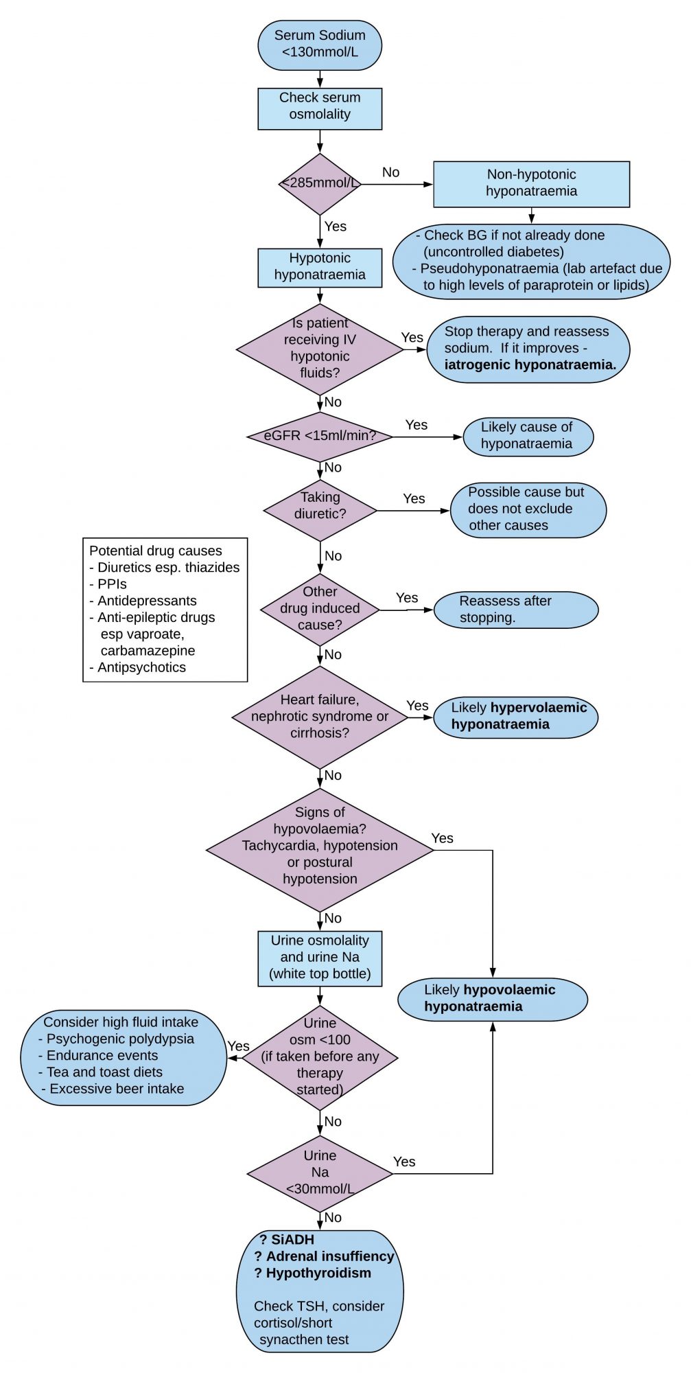
Seven Point Assessment Plan

  1. Assess biochemical severity
  2. Assess for cerebral oedema
  3. Determine duration of hyponatraemia if you can.
  4. Check serum osmolality to confirm hypo-osmolar
  5. Look for reversible causes especially drugs
  6. Assess patient’s ECF volume status
  7. Check urine sodium and osmolality

Assess Biochemical Severity

  1. Mild hyponatraemia is a biochemical finding of serum sodium 130-135 mmol/L for which obvious reversible causes should be considered but which need not generally be investigated further
  2. Serum sodium <130mmol/l is generally considered significant
  3. Symptoms/signs usually only occur when sodium <125 mmol/L.
  4. Acute hyponatraemia is less well tolerated.
  5. Aetiology is often multifactorial in medical patients

Assess for Cerebral Oedema (Hyponatraemic Encephalopathy)

  1. Seizures
  2. Coma
  3. Altered GCS

Determine Duration of Hyponatraemia if you can

  1. Those at risk of cerebral oedema have acute hyponatraemia developing within 48 hours ie brain swells because water moves across blood brain barrier before brain has had time to adapt
  2. Acute hyponatraemia defined as hyponatraemia that is documented to exist <48 hours.
  3. Hyponatraemia is chronic if documented to exist for at least 48 hours and is considered to be chronic if duration unknown, unless there is clinical evidence of the contrary

Check Serum Osmolality to Confirm Hypo-osmolar

  1. A measured serum osmolality <275 mOsm/kg always indicates hypotonic hyponatraemia.
  2. Artefactual hyponatraemia most commonly occurs when blood taken from arm into which dextrose is being infused.
  3. Can also occur in hyperlipidaemia and hyperproteinaemia (myeloma) because the aqueous fraction of the plasma specimen is reduced by the volume occupied by the lipid/protein molecules. Serum osm normal and treatment of ‘hyponatraemia’ not needed. Also known as pseudohyponatraemia.
  4. Transient hyponatraemia can also occur with acute hyperglycaemia which expands ECF by its osmotic action with fall in serum Na of 2.4 mmol/l for every 5.5 mmol/l rise in BG.

Possible Drug Induced Causes

  1. Diuretics – thiazides commonest cause of hyponatraemia  (impair free water clearance)
  2. Proton pump inhibitors
  3. Anticonvulsants esp carbamazepine and valproate
  4. Antidepressants
  5. Antipsychotics
  6. Anticancer drugs

NB All except diuretics can trigger inappropriate release of ADH

Other Reversible Causes

  1. Endocrine – hypothyroid, hypoadrenal and hypopituitary
  2. Psychogenic polydipsia ie excessive water drinking

Assess Extracellular Fluid (ECF) Volume Status

  1. Volume deplete – sodium deficit greater than water deficit.
  2. Euvolaemic – water retention only
  3. Oedematous – sodium retention with relatively greater water retention

Check Urine Sodium and Osmolality

  1. If urine Na <30mmol/l then it is likely that low effective arterial blood volume is the cause of hypotonic hyponatraemia
  2. If urine Na ≥30mmol/l then diuretics and/or renal disease likely to be responsible, failing which assess ECF volume which will either be normal eg SIADH, hypothyroidism, adrenal insufficiency; or reduced, eg cerebral salt wasting which is uncommon and usually associated with subarachnoid haemorrhage, subdural haematoma, cerebral injury or brain tumour.
  3. Urine osm is less useful than urine Na in helping to make a diagnosis
  4. If urine osm <100 then high fluid intake is the likely cause of hyponatraemia ie drinking too much water and passing large volumes dilute urine.
  5. If urine osm >700 and patient has SIADH then you are unlikely to correct hyponatraemia simply by water restriction

Causes of SIADH

  1. Malignancy esp. small cell lung cancer
  2. Primary brain injury eg. Meningitis, SAH
  3. Drugs – see list above though not all drug induced causes are due to SIADH
  4. Infections eg pneumonia
  5. Hypothyroidism

Criteria for Diagnosis of SIADH

  1. Clinically euvolaemic
  2. Low serum sodium <130mmol/l
  3. Low serum osmolality <270mosm/kg
  4. Urine Na typically >30mmol/l with normal dietary and salt intake
  5. Inappropriately concentrated urine osm >100mosm/kg
  6. No recent diuretic use – interpret biochem with caution if taking diuretic
  7. Normal serum potassium and blood urea
  8. Absence of renal, adrenal, thyroid or pituitary insufficiency

Diagnostic Difficulties with Diuretics

  1. All types of diuretics may be associated with hyponatraemia
  2. Thiazides are the most likely culprit because they impair free water clearance to a greater extent than others
  3. The use of diuretics does not exclude other causes of hyponatraemia
  4. Urine Na <30mmol/l suggests low effective arterial volume even in patients taking diuretics
  5. Urine Na >30mmol/l should be interpreted with caution if taking diuretic

Five Treatment Scenarios

  1. Hyponatraemia with Severe Symptoms (seizures, coma, altered GCS) regardless of chronicity
  2. Acute or Chronic Hyponatraemia (>48 hours or unclear duration) without Severe Symptoms
  3. Hypervolaemic Hyponatraemia
  4. Syndrome of Inappropriate Antidiuretic Hormone (SIADH)
  5. Fluid status is unclear

Hyponatraemia with Severe Symptoms (seizures, coma, altered GCS) Regardless of Chronicity

  1. Seek senior input
  2. Consider if Critical Care transfer is required for monitoring of bloods and clinical status after the infusion.  While it is safe to administer hypertonic saline on a general ward, it is essential that the subsequent monitoring and blood tests take place as outlined below; this may be easier to guarantee in a Critical Care environment.
  3. Give 150ml IV 2.7% hypertonic saline over 20 min.
  4. Check serum sodium concentration after 20 min
  5. Repeat 150ml IV 2.7% saline over 20 mins until sodium risen by 5 mmol/L
  6. Then stop 2.7% saline and give 0.9% saline 83 – 125ml/hour.
  7. Aim to limit rise in serum sodium in first 24 hours to ≤10 mmol/L and ≤8 mmol/L in each following 24 hours.
  8. Recheck sodium at 6, 12, 24 and 48 hours.

Acute or Chronic Hyponatraemia without Severe Symptoms

  1. Ensure there are no sampling or sample handling errors e.g. drip arm venepuncture.
  2. Recheck serum sodium.
  3. Stop any non-essential fluids, medications and other factors that can contribute to or provoke the hyponatraemia
  4. Make diagnostic assessment and treat underlying cause
  5. In mild hyponatraemia (Na 130-135mmol/l), there is no need to treat with the sole aim of increasing the serum sodium
  6. If hypovolaemic start IV 0.9% saline 125-250ml/hr depending on severity of fluid deficit
  7. Recheck serum sodium after 4 hours to determine trend.
  8. Aim to limit rise in serum sodium in first 24 hours to ≤ 10 mmol/L and ≤ 8mmol/L in each following 24 hours
  9. NB Risk of Central Pontine Myelinolysis if serum sodium corrected too quickly – see below
  10. Note that in case of haemodynamic instability, the need for rapid fluid resuscitation overrides the risk of an overly rapid increase in serum sodium concentration

Hypervolaemic Hyponatraemia

  1. There is insufficient data to suggest that increasing serum sodium improves patient outcomes in this group
  2. The European Clinical Practice Guideline suggests not treating simply to raise the serum sodium unless this is <125mmol/l
  3. These patients may require or already be taking a diuretic
  4. Could consider fluid restriction as a means of reducing further fluid overload

SIADH

  1. Restrict fluid intake to 1000-1200ml daily as first-line treatment in patients without severe symptoms
  2. Give combination of low dose loop diuretics and oral sodium chloride if fluid restriction alone fails. The idea here is to get rid of excess water with frusemide while increasing sodium levels with slow sodium.
  3. The dose of Slow Sodium is 4 tabs bd initially where 1 tab slow sodium contains 600mg NaCl or 10mmol Na
  4. Lithium, demeclocycline and vasopressin receptor antagonists eg tolvaptan not routinely recommended.

If Fluid Status is Unclear

  1. Give therapeutic trial of saline 0.9% 1 litre over 12 hours
  2. Recheck U&E after 6 hours
  3. Serum Na should increase if hypovolaemic
  4. Patients with SIADH don’t improve or may worsen – stop fluids if this is the case

Central Pontine Myelinolysis (CPM)

  1. This is a neurological disorder that most frequently occurs after too rapid medical correction of sodium hyponatremia.
  2. The rapid rise in sodium concentration pulls water from brain cells which leads, through a mechanism that is only partly understood, to the destruction of myelin, the substance that normally protects nerve fibers.
  3. Certain areas of the brain are particularly susceptible to myelinolysis, especially the pons.
  4. Symptoms, which begin to appear 2 to 3 days after hyponatremia is corrected, include a depressed level of awareness, dysarthria or mutism, and dysphagia.
  5. Additional symptoms often arise over the next 1-2 weeks, including impaired thinking, arm and leg weakness, stiffness, impaired sensation, and poor coordination.
  6. At its most severe, myelinolysis can lead to coma, “locked-in” syndrome (the complete paralysis of all of the voluntary muscles in the body except for those that control the eyes), and death.

Links

  1. Clinical practice guideline on diagnosis and treatment of hyponatraemia – European Journal of Endocrinology 2014; 170, G1-G47 [pdf]

Content updated by Sian Finlay